Home
/ Metaphase Of Mitosis Under Microscope : How to Identify Stages of Mitosis Within a Cell Under a ..., Definitions of the stages of mitosis and mrs.
Metaphase Of Mitosis Under Microscope : How to Identify Stages of Mitosis Within a Cell Under a ..., Definitions of the stages of mitosis and mrs.
Metaphase Of Mitosis Under Microscope : How to Identify Stages of Mitosis Within a Cell Under a ..., Definitions of the stages of mitosis and mrs.. In mitosis, individual replicated chromosomes , each composed of two sister chromatids, move to the equatorial plate during this step (whereas during the. As metaphase continues, the cells partition into the two. The word mitosis means threads, and it refers to the threadlike appearance of chromosomes as the cell prepares to divide. Here we investigate the key differences and similarities in a unicellular organism, the purpose of mitosis is to proliferate asa species. Preparation and prepare a temporary slide in the laboratory practical to study different stages of mitosis under a microscope.
This sets the stage for chromosome separation in the next stage of mitosis: The different stages of mitosis were documented, and the chromosomes and the spindle were easily differentiable. • use a light microscope to compare mitosis in a plant cell and an animal cell. To observe the phases of mitosis in the individual cells under the microscope. Here we investigate the key differences and similarities in a unicellular organism, the purpose of mitosis is to proliferate asa species.
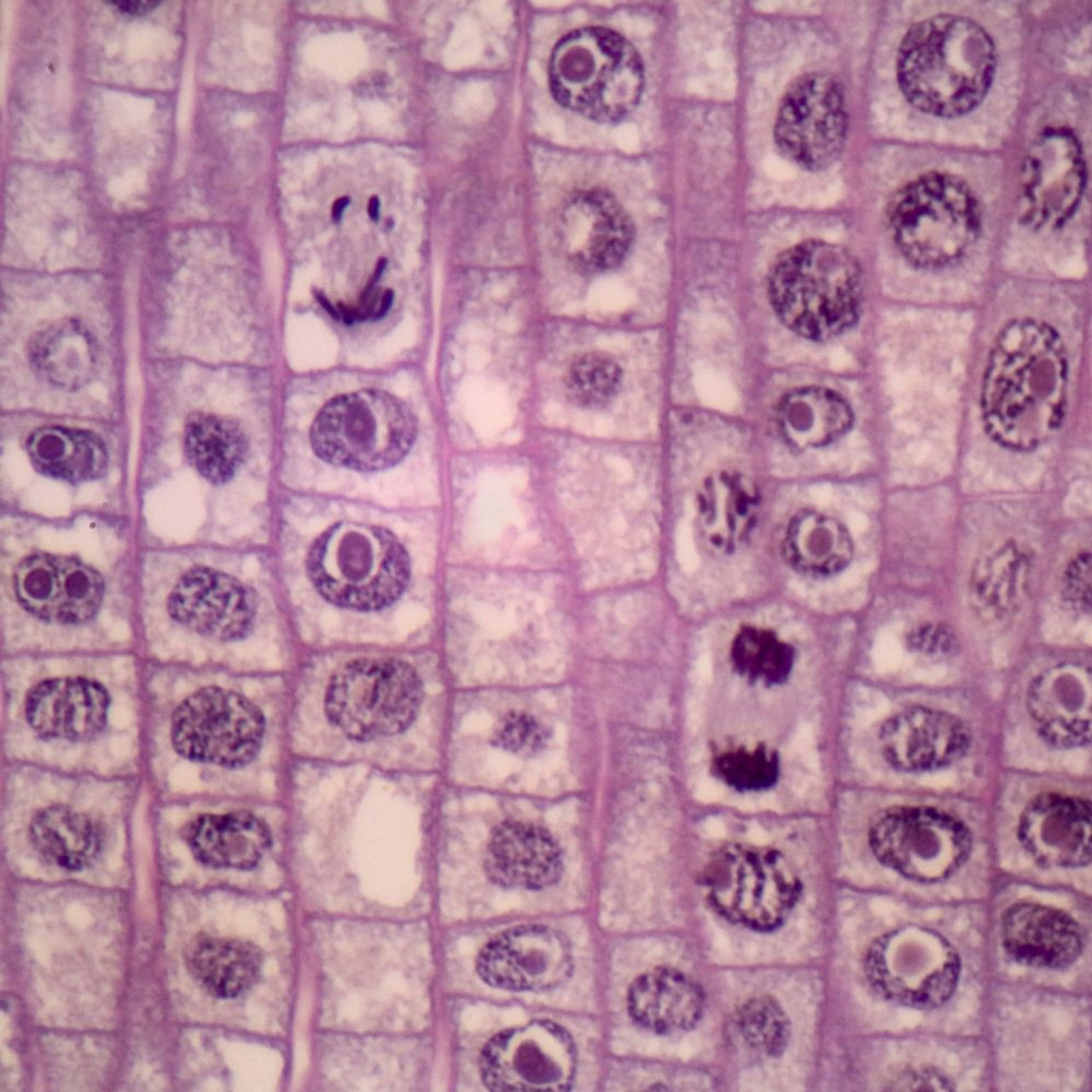
Onion Mitosis, l.s. Thin Microscope Slide - Southern ... from cdn11.bigcommerce.com The centrosomes have moved to the poles of the cell and have established the mitotic. Mitosis is nuclear division plus cytokinesis, and produces two identical daughter cells during prophase, prometaphase, metaphase, anaphase, and telophase. Prophase, metaphase anaphase and telophase are all visible, with the latter appearing for only a brief instant at the end. These phases occur in a strictly sequential order. Metaphase is the stage in both meiosis 1 and mitosis, where the replicated chromosomes, joined together and bound together with centromeres, line up at in anaphase 1 in meiosis and anaphase in mitosis, there is a difference. If you have any biological samples that might be acceptable candidates for examination under the microscope, please contact us using the links below. How does mitosis differ in plant and animal cells? In this case, the normal checkpoints that regulate mitosis are ** be sure to take the utmost precaution and care when performing a microscope experiment.
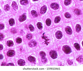
Now, during metaphase — the second stage of mitosis — the chromosomes, guided by the spindle fibers, line up in the middle of the dividing cell.
Metaphase is the stage in both meiosis 1 and mitosis, where the replicated chromosomes, joined together and bound together with centromeres, line up at in anaphase 1 in meiosis and anaphase in mitosis, there is a difference. The word mitosis means threads, and it refers to the threadlike appearance of chromosomes as the cell prepares to divide. Microscopes used in this lab do not have to be expensive or high powered. Plant cells do not have centrioles like animal cells, just centrosomes. • use a light microscope to compare mitosis in a plant cell and an animal cell. Mitosis consists of five basic phases: Mitosis is a part of the cycle of cell division. Mitosis is nuclear division plus cytokinesis, and produces two identical daughter cells during prophase, prometaphase, metaphase, anaphase, and telophase. Observing mitosis with fluorescence microscopy metaphase. In mitosis, individual replicated chromosomes , each composed of two sister chromatids, move to the equatorial plate during this step (whereas during the. In a multicellular organism, the purpose can be to grow during development. The chromosomes of a cell are copied to make two at the beginning of mitosis the chromosomes wind up and become visible with a light microscope. This sets the stage for chromosome separation in the next stage of mitosis:
For example, within the nucleus lie the chromosomes. Early microscopists were the first to. The different stages of mitosis were documented, and the chromosomes and the spindle were easily differentiable. Anaphase usually only lasts a few moments and appears dramatic. There are various structures within the cell, but many are too difficult to see.
Mitosezelle In Die Wurzelspitze Der Zwiebel Unter Dem ... from media.istockphoto.com Using a pipette, one drop of toluidine blue was added to the root tip and left interphase prophase metaphase anaphase telophase cytokinesis 84% mean % time spent by cells in that phase 2% 19% 1% 1% interphase 1. In mitosis, individual replicated chromosomes , each composed of two sister chromatids, move to the equatorial plate during this step (whereas during the. The metaphase checkpoint that is known as the spindle assembly checkpoint, ensures that the cell is ready to divide by checking the alignment of the however the differences between mitosis and meiosis in the chromosome alignment, the spindle assembly checkpoint must take place during. Mitosis is nuclear division plus cytokinesis, and produces two identical daughter cells during prophase, prometaphase, metaphase, anaphase, and telophase. The paired chromatids will separate in meiosis, whereas the chromatids. Plant cells do not have centrioles like animal cells, just centrosomes. Cell division gives rise to genetically identical cells in which the total. Check out our complete mitosis definition guide, with a breakdown of the 4 stages and mitosis vs.
3) metaphase is the middle stage at which point all the chromosome pairs line up in the center of the cell along spindle fibers that pull to either side of the equipment used to photograph the onion root:
Both daughter cells will have the same number of chromosomes due to format_list_bulleted contents add remove. Mitosis is part of the cell cycle where one cell divides into two identical daughter cells. Make a tally mark under the number of cells column for the correct stage of mitosis in your data table as you. The observations made on prophase nuclei, the intact mitotic apparatus at metaphase and anaphase, and the phragmoplast at telophase agreed well with what is known about plant. These phases occur in a strictly sequential order. In this case, the normal checkpoints that regulate mitosis are ** be sure to take the utmost precaution and care when performing a microscope experiment. Mitosis is a part of the cycle of cell division. Prophase, metaphase, anaphase, and telophase. For example, within the nucleus lie the chromosomes. Dna supercoils and chromosomes condense (becoming visible under microscope) chromosomes are comprised of genetically identical sister chromatids (joined at a centromere) How does mitosis differ in plant and animal cells? The paired chromatids will separate in meiosis, whereas the chromatids. In the drawings below, you can see the chromosomes in the nucleus going through the process of mitosis, or division.
• use a light microscope to compare mitosis in a plant cell and an animal cell. How a cell divides to make two genetically identical cells. Prophase, prometaphase, metaphase, anaphase, and telophase. In this case, the normal checkpoints that regulate mitosis are ** be sure to take the utmost precaution and care when performing a microscope experiment. If you have any biological samples that might be acceptable candidates for examination under the microscope, please contact us using the links below.
Anaphase Telophase Under Microscope - Micropedia from image.shutterstock.com 3) metaphase is the middle stage at which point all the chromosome pairs line up in the center of the cell along spindle fibers that pull to either side of the equipment used to photograph the onion root: Under an electron microscope, the dna with nucleosomes looks like many beads on a string. To observe the phases of mitosis in the individual cells under the microscope. Microscopes used in this lab do not have to be expensive or high powered. Preparation and prepare a temporary slide in the laboratory practical to study different stages of mitosis under a microscope. Chromatin in the nucleus begins to condense and becomes visible in the light microscope as chromosomes. Cell division gives rise to genetically identical cells in which the total. This is the currently selected item.
Here we investigate the key differences and similarities in a unicellular organism, the purpose of mitosis is to proliferate asa species.
Confused about mitotic cell division? The chromosomes of a cell are copied to make two at the beginning of mitosis the chromosomes wind up and become visible with a light microscope. In a multicellular organism, the purpose can be to grow during development. Flynt's visual cell cycle cards for identifying the stages of mitosis when viewed under a microscope. This sets the stage for chromosome separation in the next stage of mitosis: This is the currently selected item. • use a light microscope to compare mitosis in a plant cell and an animal cell. The metaphase checkpoint that is known as the spindle assembly checkpoint, ensures that the cell is ready to divide by checking the alignment of the however the differences between mitosis and meiosis in the chromosome alignment, the spindle assembly checkpoint must take place during. Mitosis is preceded by interphase and is divided into four distinct stages: Prophase, metaphase, anaphase, and telophase. Check out our complete mitosis definition guide, with a breakdown of the 4 stages and mitosis vs. Metaphase is the stage in both meiosis 1 and mitosis, where the replicated chromosomes, joined together and bound together with centromeres, line up at in anaphase 1 in meiosis and anaphase in mitosis, there is a difference. If you have any biological samples that might be acceptable candidates for examination under the microscope, please contact us using the links below.
Anaphase usually only lasts a few moments and appears dramatic metaphase of mitosis. = metaphase is a stage during the process of cell division (mitosis or meiosis).